
Items
Site
The Medicine Chest
keywords is exactly
Pathology Learning Centre
-

The Heterotopic Hearts
“On two occasions in 1977, when a patient’s left ventricle failed acutely after routine open-heart surgery and when no human donor organ was available, Barnard transplanted an animal heart heterotopically. On the first occasion, a baboon heart was transplanted, but this failed to support the circulation sufficiently, the patient dying some six hours after transplantation. In the second patient, a chimpanzee heart successfully maintained life until irreversible rejection occurred four days later, the recipient’s native heart having failed to recover during this period. Further attempts at xenotransplantation were abandoned and even now, more than 30 years later, xenotransplantation remains an elusive holy grail despite decades of research.” Extract: Brink JG, Hassoulas J. The first human heart transplant and further advances in cardiac transplantation at Groote Schuur Hospital and the University of Cape Town. Cardiovasc J Afr. 2009; 20(1):31-5 -

Post-mortem ledger
A wounded post-mortem ledger -

The Northern Lights
Histology slides of the brain (human and animal), scanned and converted into video projection -

Geometry in unlikely places
"These bladder stones are most likely made up of calcium oxalate; they are faceted because they rub against each other. (Faceting only occurs when there are two or more stones together)". -

Haematite Miner's Lung (Or Sidero-silicosis)
Catalogue No: R3-d55-0331. Origin: UCT Anat Path museum. Old Museum No: V:x:6. Year: not recorded. Clinical data: No further clinical or laboratory details are available other than that the patient was an emaciated 50 year old man. Macroscopy: The specimens preserved are both lungs, the heart, kidneys, spleen and and portions of liver. In the thorax, both pleural cavities were completely obliterated by a fibrous pleurisy of long-standing and both lungs were universally adherent throughout. They were stripped off with difficulty and were found to have thickening of the pleura over the upper lobe on the left side and the upper and middle lobes on the right. The lower lobes on both sides were soft and spongy while the upper lobes were dense and firm on palpation but on section there was no cavitation and no evidence of tuberculosis. The left lung showed a dense fibrosis of the whole of the upper lobe and the upper third of the lower lobe; no crepitant lung tissue could be found in the upper lobe while the lower two-thirds of the lower was crepitant and showed emphysema of a hypertrophic nature. The lung was a dull brick colour and haematite dust flowed out with the fluid when the lung was sectioned. The right lung presented a similar appearance to the left. There was a solid dense fibrosis of the upper and middle lobes and the lower lobe showed fibrosis with hypertrophic emphysema. There was no evidence of tuberculosis and on palpation, a dense fibrosis was found with no nodular formation whatever. On section, it showed a similar appearance of a brick-dust colour, dilated bronchi and uniform fibrosis of the upper and middle lobes with no crepitant lung tissue. The pericardial sac was slightly increased in size due to a hypertrophied and dilated heart. The hypertrophy was mostly on the right side and there was a terminal dilatation of the right atrium; the valves and coronary vessels unremarkable.The liver was small and on section showed venous congestion and cloudy swelling. Microscopy: On microscopy, sections of lung show a diffuse fibrosis of both upper lobes with no recognizable lung tissue. The fibrosis in areas has a slightly whorled arrangement, the centre of which is hyaline and contains no iron pigment and surrounding it is a zone of cellular tissue containing masses of iron. In the upper part of the lower lobe where the lung tissue is recognizable as such, a few nodules definitely resembling silica nodules are to be seen. In the both lower lobes a solid oedema was noted and emphysema marked. The fibrosis was not present to anything like the same extent in the lower lobes, the emphysema being the most marked feature. No evidence of tuberculosis was found in either lung, though a calcareous gland was found in the hilum. Under polarised light, the iron showed up as a golden brown with a few points of light, clear, needle-like in contra-distinction to the iron lying free in the fibrous tissue. The macrophages are beautifully shown lying inside the alveoli filled with iron dust. Percentage of Ash 16.6 Percentage of silica to ash 6.6 Percentage of silica to dry lung 1.1 Percentage of iron to ash 10.3 Percentage of iron to dry lung 6.7 Comments: In summary, the post mortem findings were of: Dense pulmonary fibrosis; hypertrophied and dilated right ventricle; failure of compensation. This condition is described as haematite miner's lung or sidero-silicosis, caused by the inhalation of dust containing silica and ferric oxide which is the principal component of the ore. The fibrosis is thought to be caused primarily by the silica and the exact role of the iron pigment in the pathogenesis of the lesion is not clear. The earliest lesions occur as small densely fibrous, sub-pleural foci usually in the upper lobes; these grow by coalescence of adjacent foci until a diffuse fibrosis of the whole lobe is produced. Haematite miner's fibrosis is commonly associated with tuberculosis and other chronic lung infections; in addition there is quite a high incidence of carcinoma of the lung reported in these cases. -

Silicosis
A gold miner using a rock drill with a water spray in an attempt to prevent the occupational disease silicosis, caused by dust inhalation. -

Lotus Leaves (Full Leaf)
"Gabriel Orozco's interest in the natural world and its relationship with humans inspired him to have actual lotus leaves etched into the printing plates, creating a man made yet true rendering of the leaf. Every vein, tear, and insect bite was captured and imprinted on special custom milled Gampi paper matching the hues of the leaf. The proofs were made as chine collé soft ground etchings, individually cut to the shape of each leaf. Orozco had a revelation while holding one of the tissue thin leaf prints up to the window: the exquisite purity that he was seeking was best displayed deconstructed and suspended in glass, illuminated all around, like a delicate specimen. The somewhat unorthodox technique used for this edition was an exciting challenge that resulted in a series of lifelike images that honors the natural world "(Lapis Press 2021). -

"I have scars on my hands from touching certain people"
"The words of Seymour Glass, eldest sibling of the Glass family – a lesser known creation of J.D. Salinger’s literary world. The story of this family of child geniuses, who appeared on a 1927 radio quiz show called 'It’s a Wise Child', is chronicled in three of his books – mostly centering on the relationship between the two eldest brothers, Buddy and Seymour. Salinger favours Seymour. He was the most brilliant and idiosyncratic of all the children and unfortunately, for the most part, only present as a memory – since he committed suicide, age 31. Buddy reminisces about one specific Seymour moment: His mother urging him to see a psychiatrist and him telling her that he doesn’t need a head doctor – he needs a hand doctor or a dermatologist, whereupon he looks at his hands and tells her he has these scars – these discolourations on them – from touching certain people. Certain heads, certain colours and textures of human hair leave permanent marks on him. When he put his hand on his little sister Franny’s head when she was still in the carriage, it left a mark. Or with her twin brother, Zooey, when he was six or seven, during a spooky movie. He put his hand on Zooey’s head when he went under the seat to avoid watching a scary scene. It left a mark. Other things, too. Charlotte, the girl he was in love with, tried to run away from him once and he grabbed her dress. It left a permanent lemon-yellow mark on the palm of his right-hand (Salinger 1964:59). His hands were filled with these discolourations – these physical manifestations of his emotions. They became a type of medical condition. And he needed to cure it" (Liebenberg 2011: 91 - 93). Salinger recounts the day of Seymour's suicide in a short story he later wrote, called 'A Perfect Day for Bananafish'. -

Neck-support
"Made from oak, the object was used to support the neck of an individual during post-mortem examinations conducted at the University of Cape Town’s mortuary unit. My first encounter with it was in Dr Yeats’s office in the Pathology Learning Centre, many years ago. Heavy in weight and invested with the traces of hundreds of individuals, it somehow seemed more representative of disease, than the 3500 specimens floating in formaldehyde displayed in the surrounding rooms " ( Liebenberg 2021: 257). -
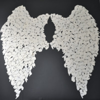
Wings
512 lasercut hands derived from images of healing: 2500 BC - 2000 AD. -
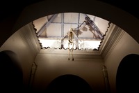
Icarus
Perspex wings with histology slides. Histology slides are prepared by taking a sample of biological tissue and fixing it to preserve the tissue in as natural a state as possible and prevent postmortem decay. The tissue is immersed in a chemical fixative and then embedded in wax to make it hard enough to cut into very thin sections of tissue (usually 5 to 7 micrometers in thickness). It is then passed through baths of solvents which remove the wax, then through graded alcohols, water and finally through baths of haematoxylin and eosin to stain it for better viewing under a microscope.
