
Items
Site
The Medicine Chest
keywords is exactly
formaldehyde
-
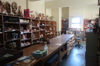
The workshop
"Held in the Department of Human Biology in the Division of Clinical Anatomy and Biological Anthropology, this collection consists of prepared specimens – both bottled and plastinated, anatomical models – and a skeletal repository used for research purposes. The materials are kept in three separate rooms, with the anatomical models in one, the prepared and plastinated specimens in another and the skeleton repository in its own room behind locked doors, viewable by appointment only. The room housing the anatomical models functioned as a workshop when modelmaking was still offered to first year medical students as an elective course: its walls are lined with shelves that display models made from materials ranging from papier-mâché to modern silicon copies and are taxonomised according to their anatomical representations, such as ‘the eye’, ‘head & neck’, ‘dentistry’, ‘embryology’, ‘lungs’ and ‘cardiovascular system’, to name a few. The models represent each anatomical section and are brightly coloured to differentiate the different parts of the human body and aid in identification. Many of the models can be dismantled into separate pieces and, like a puzzle, be reassembled into their original shapes. Interspersed with these models are rolled-up charts depicting organs, bodily systems and anatomical sections of the body in two-dimensional form, as well as old student modelling projects. A selection of animal skeletons is displayed on the far side of the room, and a shelf with a few anatomical specimens in formaldehyde is across from it" (Liebenberg 2021: 121 - 122). -
Terra Nova
Robert Falcon Scott’s Terra Nova expedition reached the South Pole on 17 January 1912 — 23 days too late. Inside a small tent supported by a single bamboo flying a Norwegian flag, was a record of the five who had been the first to reach the pole: Roald Amundsen, the leader, and his team - Olav Olavson Bjaaland, Hilmer Hanssen, Sverre H. Hassel and Oscar Wisting. On 19 January, they began their 1,300 kilometre journey home, Scott writing: “I’m afraid the return journey is going to be dreadfully tiring and monotonous” (Scott 1914: 548). -

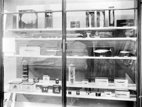
Fish
A showcase of fish found in the Artic regions. -

Haematite Miner's Lung (Or Sidero-silicosis)
Catalogue No: R3-d55-0331. Origin: UCT Anat Path museum. Old Museum No: V:x:6. Year: not recorded. Clinical data: No further clinical or laboratory details are available other than that the patient was an emaciated 50 year old man. Macroscopy: The specimens preserved are both lungs, the heart, kidneys, spleen and and portions of liver. In the thorax, both pleural cavities were completely obliterated by a fibrous pleurisy of long-standing and both lungs were universally adherent throughout. They were stripped off with difficulty and were found to have thickening of the pleura over the upper lobe on the left side and the upper and middle lobes on the right. The lower lobes on both sides were soft and spongy while the upper lobes were dense and firm on palpation but on section there was no cavitation and no evidence of tuberculosis. The left lung showed a dense fibrosis of the whole of the upper lobe and the upper third of the lower lobe; no crepitant lung tissue could be found in the upper lobe while the lower two-thirds of the lower was crepitant and showed emphysema of a hypertrophic nature. The lung was a dull brick colour and haematite dust flowed out with the fluid when the lung was sectioned. The right lung presented a similar appearance to the left. There was a solid dense fibrosis of the upper and middle lobes and the lower lobe showed fibrosis with hypertrophic emphysema. There was no evidence of tuberculosis and on palpation, a dense fibrosis was found with no nodular formation whatever. On section, it showed a similar appearance of a brick-dust colour, dilated bronchi and uniform fibrosis of the upper and middle lobes with no crepitant lung tissue. The pericardial sac was slightly increased in size due to a hypertrophied and dilated heart. The hypertrophy was mostly on the right side and there was a terminal dilatation of the right atrium; the valves and coronary vessels unremarkable.The liver was small and on section showed venous congestion and cloudy swelling. Microscopy: On microscopy, sections of lung show a diffuse fibrosis of both upper lobes with no recognizable lung tissue. The fibrosis in areas has a slightly whorled arrangement, the centre of which is hyaline and contains no iron pigment and surrounding it is a zone of cellular tissue containing masses of iron. In the upper part of the lower lobe where the lung tissue is recognizable as such, a few nodules definitely resembling silica nodules are to be seen. In the both lower lobes a solid oedema was noted and emphysema marked. The fibrosis was not present to anything like the same extent in the lower lobes, the emphysema being the most marked feature. No evidence of tuberculosis was found in either lung, though a calcareous gland was found in the hilum. Under polarised light, the iron showed up as a golden brown with a few points of light, clear, needle-like in contra-distinction to the iron lying free in the fibrous tissue. The macrophages are beautifully shown lying inside the alveoli filled with iron dust. Percentage of Ash 16.6 Percentage of silica to ash 6.6 Percentage of silica to dry lung 1.1 Percentage of iron to ash 10.3 Percentage of iron to dry lung 6.7 Comments: In summary, the post mortem findings were of: Dense pulmonary fibrosis; hypertrophied and dilated right ventricle; failure of compensation. This condition is described as haematite miner's lung or sidero-silicosis, caused by the inhalation of dust containing silica and ferric oxide which is the principal component of the ore. The fibrosis is thought to be caused primarily by the silica and the exact role of the iron pigment in the pathogenesis of the lesion is not clear. The earliest lesions occur as small densely fibrous, sub-pleural foci usually in the upper lobes; these grow by coalescence of adjacent foci until a diffuse fibrosis of the whole lobe is produced. Haematite miner's fibrosis is commonly associated with tuberculosis and other chronic lung infections; in addition there is quite a high incidence of carcinoma of the lung reported in these cases.
