
Items
Site
The Medicine Chest
keywords is exactly
heart
-
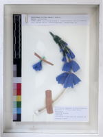
Foxgloves
Called variously foxgloves, witch’s gloves, dead men’s bells, fairy’s gloves, bloody fingers, gloves of our lady, fairy caps, virgin’s gloves or fairy thimbles, Digitalis purpurea is popular with children, who pluck the tempting bell-shaped blooms and wear them like thimbles, admonished not to lick their fingers afterwards for fear they will go blind (Young 2002: 57). While the flower can be lethal if ingested, the drug digitalis derived from foxglove is most commonly used as a heart stimulant. Digitalis has been prescribed since the 17th century, perhaps earlier, as a diuretic and to slow the pulse and is still the drug of choice for atrial fibrillation. In the 1770s, William Withering got an old family recipe, a herbal infusion for treating swollen legs, from a Shropshire woman and identified digitalis as the active one of the twenty ingredients. Many more antiarrhythmic drugs now exist (Young 2002: 57). Made up of surgical gloves, Band-Aids, syringes and IV-tubing (with an infusion of foxglove leaves in its stem) the work mimicked the language of the herbarium specimen, drawing a viewer in to examine the content they were expecting, only to surprise them with its incongruous materials. -
The Mother of all Firewalls
'The Mother of all Firewalls' (2012), is a sculptural piece by Kim Gurney made from beeswax, gold glitter, bitumen, repurposed tiles, rabbit skin and glue, which seems to depict an EEG graph in its format but in reality references a graph plotted by Google Insights after Gurney inputted a range of new words that emerged from the ‘Eurogeddon’ of 2012, showing their incidence in news reports pertaining to this financial crisis between 2008 and 2012. -
Subtle Thresholds (PLC)
"Integrated into the PLC, these works speak to the specimens on display and provide interesting access points to the collection. The animal-faeces prints (which referenced ‘sites of contamination’ in the context of the SAM exhibition) resonate, for instance, with many of the specimens on display in the PLC, such as the heterotopic heart (also called a ‘piggy-back heart transplant’) created by Dr Chris Barnard in 1977, consisting of a baboon heart grafted onto a human heart for additional motoric support. In addition to their more ‘famous’ specimens, the centre also has an extensive intestinal worm collection and many organs affected by zoonotic diseases, such as a liver ravaged by malaria. This was not a conscious decision on the part of the artist-curator and illustrates how curation can draw attention to aspects of a collection and liberate new associations when brought into conversation with it" (Liebenberg 2021: 201). -

Tabloid medicine chest from Scott Polar Expedition
The expedition carried with them a Tabloid Medicine Chest. On the 11th of March, knowing that the party was unlikely to survive, Scott ordered Edward Wilson, the expedition’s Chief Scientist and a qualified doctor, to divide the painkillers between them so they could each end their life on their own terms. Writing in his diary on that day, Scott states, “I practically ordered Wilson to hand over the means of ending our troubles to us, so that anyone of us may know how to do so". Scott, Bowers and Oates had thirty opium tabloids apiece and Wilson, the morphine. Scotts diary entry on either the 22nd or 23rd of March showed that they had a change of heart however: “no fuel and only one or two left of food — must be near the end. Have decided that it shall be natural — we shall march for the depot with or without our effects and die in our tracks". -

The Heterotopic Hearts
“On two occasions in 1977, when a patient’s left ventricle failed acutely after routine open-heart surgery and when no human donor organ was available, Barnard transplanted an animal heart heterotopically. On the first occasion, a baboon heart was transplanted, but this failed to support the circulation sufficiently, the patient dying some six hours after transplantation. In the second patient, a chimpanzee heart successfully maintained life until irreversible rejection occurred four days later, the recipient’s native heart having failed to recover during this period. Further attempts at xenotransplantation were abandoned and even now, more than 30 years later, xenotransplantation remains an elusive holy grail despite decades of research.” Extract: Brink JG, Hassoulas J. The first human heart transplant and further advances in cardiac transplantation at Groote Schuur Hospital and the University of Cape Town. Cardiovasc J Afr. 2009; 20(1):31-5 -

A beating heart
"Dear Doctor Barnard I am an 11 year old girl, and I have a problem: I went fishing today; when we came back, my parents cleaned the fish, and after they took out the insides, they found a heart of a fish beating, but the fish was dead and cut up. It was still beating for about 1/2 an hour. Can you explain that? I am very interested in biology, and so is everyone else in my family. Sincerely Yours, Lillian Levy P.S. I know you are a very busy man, but, if you have enough time, please try to answer. THANK YOU! P.P.S. If a doctor says you're dead and they take out your heart but it is still beating, are you dead or alive?" Transcribed letter from the Heart of Cape Town Museum -

Haematite Miner's Lung (Or Sidero-silicosis)
Catalogue No: R3-d55-0331. Origin: UCT Anat Path museum. Old Museum No: V:x:6. Year: not recorded. Clinical data: No further clinical or laboratory details are available other than that the patient was an emaciated 50 year old man. Macroscopy: The specimens preserved are both lungs, the heart, kidneys, spleen and and portions of liver. In the thorax, both pleural cavities were completely obliterated by a fibrous pleurisy of long-standing and both lungs were universally adherent throughout. They were stripped off with difficulty and were found to have thickening of the pleura over the upper lobe on the left side and the upper and middle lobes on the right. The lower lobes on both sides were soft and spongy while the upper lobes were dense and firm on palpation but on section there was no cavitation and no evidence of tuberculosis. The left lung showed a dense fibrosis of the whole of the upper lobe and the upper third of the lower lobe; no crepitant lung tissue could be found in the upper lobe while the lower two-thirds of the lower was crepitant and showed emphysema of a hypertrophic nature. The lung was a dull brick colour and haematite dust flowed out with the fluid when the lung was sectioned. The right lung presented a similar appearance to the left. There was a solid dense fibrosis of the upper and middle lobes and the lower lobe showed fibrosis with hypertrophic emphysema. There was no evidence of tuberculosis and on palpation, a dense fibrosis was found with no nodular formation whatever. On section, it showed a similar appearance of a brick-dust colour, dilated bronchi and uniform fibrosis of the upper and middle lobes with no crepitant lung tissue. The pericardial sac was slightly increased in size due to a hypertrophied and dilated heart. The hypertrophy was mostly on the right side and there was a terminal dilatation of the right atrium; the valves and coronary vessels unremarkable.The liver was small and on section showed venous congestion and cloudy swelling. Microscopy: On microscopy, sections of lung show a diffuse fibrosis of both upper lobes with no recognizable lung tissue. The fibrosis in areas has a slightly whorled arrangement, the centre of which is hyaline and contains no iron pigment and surrounding it is a zone of cellular tissue containing masses of iron. In the upper part of the lower lobe where the lung tissue is recognizable as such, a few nodules definitely resembling silica nodules are to be seen. In the both lower lobes a solid oedema was noted and emphysema marked. The fibrosis was not present to anything like the same extent in the lower lobes, the emphysema being the most marked feature. No evidence of tuberculosis was found in either lung, though a calcareous gland was found in the hilum. Under polarised light, the iron showed up as a golden brown with a few points of light, clear, needle-like in contra-distinction to the iron lying free in the fibrous tissue. The macrophages are beautifully shown lying inside the alveoli filled with iron dust. Percentage of Ash 16.6 Percentage of silica to ash 6.6 Percentage of silica to dry lung 1.1 Percentage of iron to ash 10.3 Percentage of iron to dry lung 6.7 Comments: In summary, the post mortem findings were of: Dense pulmonary fibrosis; hypertrophied and dilated right ventricle; failure of compensation. This condition is described as haematite miner's lung or sidero-silicosis, caused by the inhalation of dust containing silica and ferric oxide which is the principal component of the ore. The fibrosis is thought to be caused primarily by the silica and the exact role of the iron pigment in the pathogenesis of the lesion is not clear. The earliest lesions occur as small densely fibrous, sub-pleural foci usually in the upper lobes; these grow by coalescence of adjacent foci until a diffuse fibrosis of the whole lobe is produced. Haematite miner's fibrosis is commonly associated with tuberculosis and other chronic lung infections; in addition there is quite a high incidence of carcinoma of the lung reported in these cases. -

Lacuna (Part one)
"It is interesting to note that the botanical origins of most of these medicines were from outside of Africa, especially if one considers the long history of the Cape as a point on the trade routes where ill sailors regularly disembarked and drew on the knowledge of the Khoekhoe traditional healers for treatment and herbal cures (Laidler & Gelfand 1971: 44). The Cape flora offered a plenitude of medicinal resources and these healers (who were skilled in botany, surgery and medicine) used them in a variety of healing practices . The exclusion of local botanical remedies in the BWC No. 254 medicine chest can be attributed to many factors" (Liebenberg 2021: 67). -
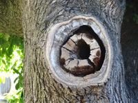
Lacuna (Part two)
An ill English Oak on Hiddingh Campus, Michaelis School of Fine Art, University of Cape Town. English oaks were first brought into the country by the early settlers and were one of the first exotic tree species to be planted in South Africa, shortly after Van Riebeek’s arrival in 1652. He explained that in South Africa, these trees do not grow as old as they would have in Europe. The high temperatures cause these trees to grow faster than their species back home, and because of this, their centers start rotting over an extended period of time. The center part of the wood – the heart – is affected by this occurrence and hollowed out over time. -
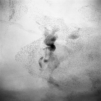
Murmeration
Heart murmurs are sounds – such as whooshing or swishing – made by turbulent blood in or near the heart. When doctors listen to a child's heart, what they usually hear is a simple rhythm: "lub-dub, lub-dub, lub-dub..." Sometimes, they'll hear an extra sound in between the lub and the dub. That extra sound is called a heart murmur. Heart murmurs can be harmless or abnormal. In the case of the latter, it is usually the result of abnormal blood flow through the heart caused by a heart valve not working properly. -

Murmeration
A short film that follows the journey of two girls in a canoe on the River Shannon and how they stumble across one of nature's greatest phenomenons; a murmuration of starlings.
