
Items
Site
The Medicine Chest
keywords is exactly
patient
-
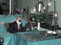
Dogs in the Heart of Cape Town
The guided tour of the museum, which commemorates the first heart transplant performed by Chris Barnard in 1967, starts with a representation of the car accident that provided the heart for the transplant, through to the animal lab where Barnard conducted experiments with over 50 dogs to perfect the technique of heart transplantation. From there one can tour a model of Denise Darvall's bedroom and Christiaan Barnard's office before seeing a recreation of the surgery in the actual operating theaters where it occurred. -

Haematite Miner's Lung (Or Sidero-silicosis)
Catalogue No: R3-d55-0331. Origin: UCT Anat Path museum. Old Museum No: V:x:6. Year: not recorded. Clinical data: No further clinical or laboratory details are available other than that the patient was an emaciated 50 year old man. Macroscopy: The specimens preserved are both lungs, the heart, kidneys, spleen and and portions of liver. In the thorax, both pleural cavities were completely obliterated by a fibrous pleurisy of long-standing and both lungs were universally adherent throughout. They were stripped off with difficulty and were found to have thickening of the pleura over the upper lobe on the left side and the upper and middle lobes on the right. The lower lobes on both sides were soft and spongy while the upper lobes were dense and firm on palpation but on section there was no cavitation and no evidence of tuberculosis. The left lung showed a dense fibrosis of the whole of the upper lobe and the upper third of the lower lobe; no crepitant lung tissue could be found in the upper lobe while the lower two-thirds of the lower was crepitant and showed emphysema of a hypertrophic nature. The lung was a dull brick colour and haematite dust flowed out with the fluid when the lung was sectioned. The right lung presented a similar appearance to the left. There was a solid dense fibrosis of the upper and middle lobes and the lower lobe showed fibrosis with hypertrophic emphysema. There was no evidence of tuberculosis and on palpation, a dense fibrosis was found with no nodular formation whatever. On section, it showed a similar appearance of a brick-dust colour, dilated bronchi and uniform fibrosis of the upper and middle lobes with no crepitant lung tissue. The pericardial sac was slightly increased in size due to a hypertrophied and dilated heart. The hypertrophy was mostly on the right side and there was a terminal dilatation of the right atrium; the valves and coronary vessels unremarkable.The liver was small and on section showed venous congestion and cloudy swelling. Microscopy: On microscopy, sections of lung show a diffuse fibrosis of both upper lobes with no recognizable lung tissue. The fibrosis in areas has a slightly whorled arrangement, the centre of which is hyaline and contains no iron pigment and surrounding it is a zone of cellular tissue containing masses of iron. In the upper part of the lower lobe where the lung tissue is recognizable as such, a few nodules definitely resembling silica nodules are to be seen. In the both lower lobes a solid oedema was noted and emphysema marked. The fibrosis was not present to anything like the same extent in the lower lobes, the emphysema being the most marked feature. No evidence of tuberculosis was found in either lung, though a calcareous gland was found in the hilum. Under polarised light, the iron showed up as a golden brown with a few points of light, clear, needle-like in contra-distinction to the iron lying free in the fibrous tissue. The macrophages are beautifully shown lying inside the alveoli filled with iron dust. Percentage of Ash 16.6 Percentage of silica to ash 6.6 Percentage of silica to dry lung 1.1 Percentage of iron to ash 10.3 Percentage of iron to dry lung 6.7 Comments: In summary, the post mortem findings were of: Dense pulmonary fibrosis; hypertrophied and dilated right ventricle; failure of compensation. This condition is described as haematite miner's lung or sidero-silicosis, caused by the inhalation of dust containing silica and ferric oxide which is the principal component of the ore. The fibrosis is thought to be caused primarily by the silica and the exact role of the iron pigment in the pathogenesis of the lesion is not clear. The earliest lesions occur as small densely fibrous, sub-pleural foci usually in the upper lobes; these grow by coalescence of adjacent foci until a diffuse fibrosis of the whole lobe is produced. Haematite miner's fibrosis is commonly associated with tuberculosis and other chronic lung infections; in addition there is quite a high incidence of carcinoma of the lung reported in these cases. -
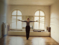
Touching both walls at the same time
As a young sculptor working in the 1960s, Horn suffered severe lung damage from working with fiberglass and polyester, and spent long periods convalescing in hospital. Whilst restricted to her hospital bed, Horn devised a series of wearable sculptures or 'body extensions' which she would later make using cloth, wood, bandages, belts, feathers, and found objects. Her masks and extensions contain, constrain, and/or elongate the bodies of their wearers. -
Denise Darvall
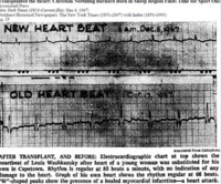
"During the first heart transplant a shift occurred in the heart of Denise Darvall, the young brain-dead car accident victim whose family had allowed her heart to be given up. In his account of the operation, Barnard writes how her heart’s life fluid returned from the lungs – how many million times had it happened? – but different this time, void of oxygen. How her heart would react, at first, as if meeting only a small inconvenience. Unaware of what was happening, it would simply pump more excitedly – expecting some relief. Yet this would never come, and it would fall back in the first wave of confusion and fatigue. Barnard equates Darvall's heart with a bird trying to take flight" (Liebenberg 2011: 107-108). -

Touching both walls at the same time
As a young sculptor working in the 1960s, Horn suffered severe lung damage from working with fiberglass and polyester, and spent long periods convalescing in hospital. Whilst restricted to her hospital bed, Horn devised a series of wearable sculptures or 'body extensions' which she would later make using cloth, wood, bandages, belts, feathers, and found objects. Her masks and extensions contain, constrain, and/or elongate the bodies of their wearers.
