
Items
Site
The Medicine Chest
keywords is exactly
teaching
-

Kimberlite
A display outside the Cape Town Diamond Museum in the V&A Waterfront -

Kimberlite
Kimberlite specimens, UCT Mantle Room -

Kimberlite
Kimberlite specimen, UCT Mantle Room -

The Diamond Mines of South Africa: Some Account of their Rise and Development.
“In the mines operated by the De Beers Company alone, more than eleven thousand African natives are employed below and above ground, coming from the Transvaal, Basutoland, and Bechuanaland, from districts far north of the Limpopo and the Zambesi, and from the Cape Colony on the east and the south to meet the swarms flocking from Delagoa Bay and countries along the coast of the Indian Ocean, while a few cross the continent from Damaraland and Namaqualand, and the coast washed by the Atlantic. The larger number are roughly classed as Basutos, Shanganes, M'umbanes, and Zulus, but there are many Batlapins from Bechuanaland, Amafengu, and a sprinkling of nearly every other tribe in South Africa” (Williams 1902: 412-413). -

The Diamond Mines of South Africa: Some Account of their Rise and Development.
“The initial impetus for establishing a collection of mantle materials for research purposes in South Africa was provided by Gardner Williams and his son Alpheus Williams in the late 19th and early 20th century. These two American mining engineers shared a great interest in the mining methods and geology of the South African kimberlite-hosted diamond mines they were supervising. Each wrote books on the subject and the two men assembled a collection of scientifically interesting rock samples and minerals from the mines. Subsequent to the death of Alpheus Williams, the Williams family donated this collection to the Geology Department at UCT for teaching and research purposes” (Department of Geological Sciences 2021). -
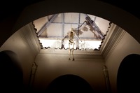
Icarus
Perspex wings with histology slides. Histology slides are prepared by taking a sample of biological tissue and fixing it to preserve the tissue in as natural a state as possible and prevent postmortem decay. The tissue is immersed in a chemical fixative and then embedded in wax to make it hard enough to cut into very thin sections of tissue (usually 5 to 7 micrometers in thickness). It is then passed through baths of solvents which remove the wax, then through graded alcohols, water and finally through baths of haematoxylin and eosin to stain it for better viewing under a microscope.
